
Managing an autoimmune disease isn’t just about taking medication. It’s about catching changes before they become crises. That’s where autoimmune disease monitoring comes in - a steady, smart system that tracks your body’s signals through labs, scans, and regular check-ins. Without it, flares sneak up. Organs get damaged. Quality of life drops. But with the right monitoring, many people avoid hospital visits, slow progression, and stay in control.
What Lab Tests Actually Tell You
Not all blood tests are created equal when it comes to autoimmune diseases. Some give you a snapshot. Others tell you if things are getting worse - or better.
The most common starter test is the ANA (antinuclear antibody). It’s positive in 95% of people with systemic lupus, but here’s the catch: up to 20% of healthy people also test positive. That’s why ANA alone doesn’t diagnose or track disease activity. A positive result just means you need more tests.
That’s where reflex testing comes in. If ANA is positive, labs run an ENA panel - looking for specific antibodies like SS-A, SS-B, Scl-70, and Jo-1. These help pinpoint the exact condition. For example, SS-A shows up in over 80% of Sjögren’s cases. Scl-70 is a red flag for systemic sclerosis. Jo-1 points to polymyositis.
Then there’s anti-dsDNA. This one’s gold for lupus. It’s 95% specific - meaning if it’s high, lupus is likely active, especially if kidney involvement is suspected. But it’s only positive in 60-70% of lupus patients, so a normal result doesn’t rule out flares.
Don’t waste time tracking ANA levels over time. They don’t change with disease activity. Instead, watch complement levels - C3 and C4. When they drop, it means your immune system is burning hot. That’s a real signal of active lupus, not just a lab curiosity.
CRP and ESR are your inflammation thermometers. CRP above 3.0 mg/L? That’s active inflammation. ESR over 20 mm/hr in women or 15 mm/hr in men? Also a red flag. These aren’t perfect - they can rise with infection or even stress - but when they climb together, it’s a strong clue your autoimmune condition is stirring.
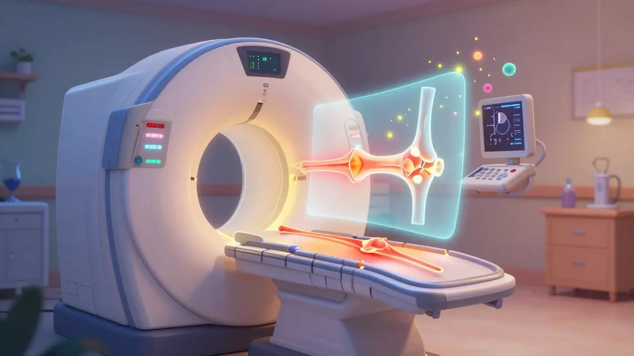
Imaging: Seeing What Blood Tests Can’t
Lab tests show what’s happening in your blood. Imaging shows what’s happening inside your joints, organs, and tissues - often before you feel pain.
MRI is the go-to for early inflammation. It can spot swelling in the synovium (joint lining) or brain lesions in lupus or MS long before X-rays show damage. New nanotech contrast agents are replacing old gadolinium dyes, reducing kidney risks and giving clearer pictures.
Ultrasound with microbubble contrast is changing how rheumatoid arthritis is tracked. It measures blood flow in inflamed joints with 85% accuracy. No radiation. No needles. Just a quick scan that shows if your treatment is calming the inflammation or not.
PET scans are newer but powerful. By tagging immune cells with radioactive tracers, doctors can see where T-cells are gathering - like a heat map of your autoimmune attack. Total-body PET, once only for cancer, now shows immune activity across the whole body in one scan.
SPECT uses radiolabeled peptides to track specific molecules at inflammation sites. It’s not as common, but in complex cases - like vasculitis or unexplained organ damage - it can reveal hidden hotspots.
CT scans give detailed structure. They’re great for spotting lung scarring in scleroderma or bowel thickening in Crohn’s. But they use radiation, so they’re not used for frequent monitoring.
How Often Should You See Your Doctor?
There’s no one-size-fits-all schedule. It depends on how active your disease is and which organs are involved.
If you’ve just been diagnosed or your symptoms are flaring, expect visits every 4 to 6 weeks. That’s when doctors adjust meds, check labs, and decide if you need imaging.
Once you’re stable - meaning no new symptoms, normal labs, and no new damage on scans - visits drop to every 3 to 4 months. The American College of Rheumatology says you still need at least two full checkups a year, even if you feel fine.
For high-risk patients - those with kidney, lung, or brain involvement - quarterly visits with full labs and imaging are standard. If you only have joint pain and mild fatigue, and your disease has been quiet for two years, you might stretch to 6-12 months between visits.
Guidelines from EULAR and ACR use disease activity scores to guide this. For rheumatoid arthritis, they use DAS28. For lupus, it’s SLEDAI. These aren’t just fancy numbers - they combine how you feel, physical exam findings, and lab results into one score. If your score goes up, your treatment needs a tweak.
What Doctors Actually Weigh
Here’s the truth: no single test tells the whole story. Experts at the 2023 International Autoimmune Summit broke it down:
- 30% - Lab markers (CRP, ESR, antibodies, complements)
- 30% - Imaging findings (MRI, ultrasound, PET)
- 40% - Clinical assessment (how you feel, what your doctor sees)
Dr. Betty Hahn from UNC put it bluntly: “Relying only on lab values misses critical clinical context in 63% of flares.”
That means if your CRP is normal but your knees are swollen and you’re exhausted, you’re not fine. If your MRI shows joint damage but you feel great and your labs are clean, you might still need treatment. The human piece - your symptoms, your energy, your pain - carries the most weight.
New Tech on the Horizon
Monitoring isn’t stuck in the past. Wearables are starting to pick up inflammatory signals through interstitial fluid - the fluid between your cells. Early studies show these devices match traditional CRP readings 89% of the time. Imagine getting a notification on your watch that inflammation is rising - before you even feel it.
AI is stepping in too. Platforms like AutoimmuneTrack, approved by the FDA in 2023, analyze your lab history, symptom logs, and wearable data to predict flares 14 days in advance with 76% accuracy. In a trial of over 2,300 patients, emergency visits dropped by 29%.
CyTOF (mass cytometry) is another leap. Instead of measuring 15 immune cell types at once like old flow cytometry, it can track 50. That means seeing rare immune cells that drive flares - and targeting them better.
But these tools aren’t for everyone. Cost, access, and insurance hurdles are real. Only 48% of Medicaid patients get recommended monitoring, compared to 83% of those with private insurance. And test results vary by lab - ANA results can differ by 22% between facilities. That’s why it’s best to stick with the same lab and doctor over time.

What to Ask Your Doctor
Don’t wait for your appointment to wonder what’s going on. Bring these questions:
- Which lab markers are most important for my specific disease?
- Are my current test results trending up, down, or staying the same?
- Do I need imaging this visit? Why or why not?
- What’s my disease activity score? Has it changed since last time?
- Are my symptoms matching my lab results? If not, why?
- What’s the plan if my numbers worsen?
Monitoring isn’t about fear. It’s about power. The more you know, the more you can work with your team to stay ahead of your disease - not react to it.
Is ANA testing useful for tracking autoimmune disease over time?
No. ANA levels often stay positive even when the disease is in remission. Tracking them over time doesn’t tell you if your condition is flaring or improving. Instead, focus on complement levels (C3 and C4), CRP, ESR, and disease-specific antibodies like anti-dsDNA for lupus. These change with activity and give real insight into what’s happening in your body.
Why do I need imaging if my blood tests are normal?
Blood tests show inflammation in your bloodstream, but they can’t see damage inside your joints, organs, or tissues. MRI and ultrasound can detect early swelling or structural changes before you feel pain or before labs show abnormalities. For example, joint damage in rheumatoid arthritis often starts silently - imaging catches it early, so treatment can prevent permanent harm.
How often should I get blood work done for autoimmune disease monitoring?
If your disease is active or you’re starting a new treatment, expect blood tests every 4-6 weeks. Once stable, most people need testing every 3-4 months. Even if you feel fine, at least two comprehensive lab checks per year are recommended. Your doctor will adjust based on your disease type, severity, and response to treatment.
Can wearable devices replace lab tests for autoimmune monitoring?
Not yet. Wearables that track inflammatory signals through skin fluid are promising and show strong correlation with CRP levels - about 89% accuracy. But they’re still early-stage tools. They can alert you to possible changes, but they don’t replace lab tests for autoantibodies, complements, or organ function. Think of them as early warning systems, not diagnostics.
Why is my doctor ordering a PET scan for my autoimmune disease?
PET scans are used when standard tests don’t explain your symptoms or when doctors suspect widespread immune activity. They use radioactive tracers to show where immune cells are clustered - like finding hidden inflammation in the lungs, gut, or brain. This is especially helpful in complex cases like vasculitis, sarcoidosis, or unexplained multi-organ involvement. It’s not routine, but it can change your treatment path.
What if I can’t afford recommended imaging or lab tests?
Cost is a real barrier - only 48% of Medicaid patients get full monitoring compared to 83% of those with private insurance. Talk to your doctor about alternatives. Some tests can be spaced out if your condition is stable. Ask about financial aid programs at hospitals or labs. Some research centers offer free monitoring for eligible patients. Don’t skip care because of cost - work with your team to find a realistic plan.
Next Steps: Building Your Monitoring Plan
Start by writing down your last three lab results - CRP, ESR, complement levels, and key antibodies. Note when you last had an MRI, ultrasound, or other scan. Track your symptoms: fatigue, joint pain, rashes, fever - and when they happen.
At your next visit, ask: “What’s my current disease activity score? What’s the goal for next month? What test or scan will tell me if we’re on track?”
Don’t wait for a crisis. The best outcomes come from people who monitor like they’re coaching a team - consistently, calmly, and with clear goals. Your body gives you signals. Learning how to read them is the most powerful tool you have.
Selina Warren
January 17, 2026
Finally, someone gets it. I’ve been screaming this for years-ANA is useless for tracking. My doc kept chasing it like it was a crystal ball while my C3/C4 crashed and I was barely walking. When we switched to watching complements and CRP? My flares dropped 70%. Stop wasting time on antiquated markers. Your body doesn’t care about your lab’s old-school checklist.
christian Espinola
January 18, 2026
Let me guess-this was written by a pharma rep. ANA is a red herring? Sure. And the sun doesn’t rise in the east. They’re all just pushing ‘reflex panels’ because they make more money. And don’t get me started on PET scans-radiation is just a Trojan horse for tracking your biologics compliance. The FDA approves everything that keeps the machine running. You’re being played.
Chuck Dickson
January 19, 2026
Man, this is exactly what I needed to hear. I’ve been feeling like a lab rat-same blood draws, same questions, no answers. But this? This makes sense. I started tracking my CRP and ESR on my phone app, and I caught a flare two weeks before my knee swelled up. I didn’t even know I was getting sick. Monitoring isn’t paranoia-it’s power. If you’re not doing this, you’re flying blind.
Pat Dean
January 21, 2026
Typical liberal medical propaganda. You people think science is just ‘trending’ and ‘data’ and ‘wearables.’ Meanwhile, real medicine is being replaced by tech bros with apps. My grandma survived lupus with aspirin and prayer. Now you’re getting PET scans because you’re too lazy to ‘listen to your body.’
Jay Clarke
January 22, 2026
Okay, but let’s be real-how many of you are getting these tests because you’re actually sick, or because your doctor’s office is just trying to hit their KPIs? I’ve had three MRIs in six months and zero improvement. They’re not treating me-they’re billing. And now they want me to wear a watch that ‘predicts flares’? Next they’ll sell me a subscription to ‘AutoimmuneTrack Premium’ so I can get notified when my immune system hates me.
Robert Davis
January 23, 2026
Interesting. I’ve been reading up on this. The thing nobody talks about is how much variability there is between labs. My ANA was positive at Lab A, negative at Lab B. Same blood. Same day. I asked the tech-she said ‘different reagents.’ So if your ‘gold standard’ marker changes based on which tube you use… what’s even the point?
Eric Gebeke
January 23, 2026
You all sound like you’re trying to sell this to someone who doesn’t have insurance. I’ve been on Medicaid for 8 years. I get one blood test a year. They won’t cover ultrasound. They won’t cover MRI unless I’m in the ER. And now you’re telling me I need a PET scan? That’s not monitoring. That’s a luxury. And the fact that you’re talking about it like it’s normal? That’s the real disease.
Jake Moore
January 23, 2026
Just wanted to add-wearables aren’t replacing labs, but they’re giving you daily data that labs can’t. I’ve got a continuous monitor that tracks interstitial fluid cytokines. It spiked two days before my fatigue hit. I called my rheum, got a steroid pack early, and skipped the ER. That’s not magic. That’s prevention. You don’t need to be rich to use this-some clinics give them out for free if you’re in a trial.
Joni O
January 23, 2026
thank you for this. i’ve been trying to explain this to my dr for months but she just says ‘we’ll check your ana next visit.’ i started keeping a symptom journal with my crp and esr numbers and honestly? it changed everything. i finally feel like i’m part of the team, not just a patient waiting for the next crisis. also, i’m not great at spelling but i hope this makes sense lol
Ryan Otto
January 23, 2026
How quaint. You believe in Western allopathic dogma. The true cause of autoimmune disease is electromagnetic poisoning from 5G towers and glyphosate-laced vaccines. The entire lab system is a construct designed to keep you dependent on pharmaceuticals. PET scans? They’re not detecting inflammation-they’re mapping your neural signature for government surveillance. The real monitoring is spiritual. Meditation reduces CRP more than biologics ever could.
Max Sinclair
January 24, 2026
I really appreciate how clear this is. I’ve had RA for 12 years and I’ve always felt guilty for asking for imaging ‘just because.’ But now I get it-it’s not about being anxious. It’s about being proactive. My doc finally agreed to an ultrasound last month and found synovitis I didn’t even know I had. I’m not scared of monitoring anymore. I’m grateful for it.
Praseetha Pn
January 26, 2026
OMG this is so true!! I’m from India and my doctor here says ‘ANA is enough’ and refuses to do ENA or C3/C4 because ‘it’s too expensive.’ But I paid out of pocket for my own tests and my Jo-1 was positive-turns out I have polymyositis, not just ‘fatigue.’ Now I’m on methotrexate and I can actually lift my arms again. Don’t let your doctor’s laziness kill you. Fight for your data. Your body is not a mystery-it’s a puzzle you deserve to solve.
Nishant Sonuley
January 28, 2026
Look, I get it. You want to be the ‘smart patient.’ You track your CRP like it’s your fantasy football stats. But here’s the irony-you’re still relying on a system that treats you like a data point. The real solution isn’t more scans or wearables. It’s systemic change. Why are we even talking about ‘monitoring’ instead of curing? Why are we stuck in this loop of managing symptoms while Big Pharma rakes in billions? You’re optimizing the cage, not escaping it. And that’s… kinda sad.
Emma #########
January 29, 2026
This made me cry a little. I’ve been silent about my lupus for years because I didn’t want to be ‘that person.’ But reading this? I finally feel seen. I started writing down my symptoms every morning. I didn’t realize how much my energy dropped on days my ESR spiked. I’m not fixed. But I’m not invisible anymore.
Andrew McLarren
January 30, 2026
While the information presented is both comprehensive and generally aligned with contemporary clinical guidelines, it is imperative to underscore the necessity of individualized patient management. The heterogeneity of autoimmune pathophysiology necessitates a nuanced, case-specific approach to biomarker interpretation and imaging utilization. Furthermore, the socioeconomic disparities in access to advanced diagnostic modalities remain a significant ethical concern within the healthcare ecosystem.
Selina Warren
January 30, 2026
Andrew, your comment is technically correct. But no one’s here for a medical journal. We’re here because we’re tired of being treated like a spreadsheet. If your doctor can’t explain why your C4 dropped from 22 to 8 in three weeks, then your ‘nuanced approach’ is just a fancy way of saying ‘I don’t know what’s going on.’